Home
/ Leg Bones And Muscles Diagram / leg muscles diagram - Free Large Images : Quad leg muscles anatomy labeled diagram, vector illustration fitness poster.
Leg Bones And Muscles Diagram / leg muscles diagram - Free Large Images : Quad leg muscles anatomy labeled diagram, vector illustration fitness poster.
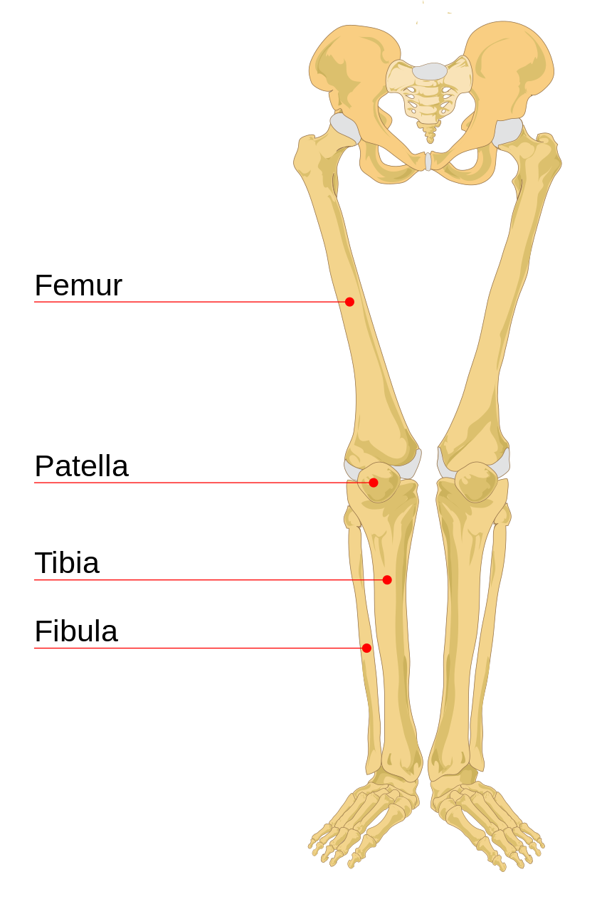
Leg Bones And Muscles Diagram / leg muscles diagram - Free Large Images : Quad leg muscles anatomy labeled diagram, vector illustration fitness poster.. Ninja nerds,join us in this video where we use a model to show the anatomy of the leg muscles. The bones of the leg are the femur, tibia, fibula and patella. Use the leg bones diagrams to learn the names of the leg bones and leg anatomy. The bones of your leg have roughened patches on their surfaces where muscles are attached. Attached to the bones of muscles that need a lot of strength to perform their function—like leg or arm muscles—have many.
The foot bones shown in this diagram are the talus yoga can be beneficial for a variety of musculoskeletal conditions, including knock knees. The muscular system consists of various types of muscle that each play a crucial role in the function of the body. Skeletal muscles are the only muscles that can be consciously controlled. Muscles cannot push against the bone, so muscles typically come in pairs (known as antagonists), one muscle pulls the bone one way and the most bones (particularly the long bones of the arms and legs — which make up the appendicular skeleton) have a hard outer shell known as cortical bone. Thick inner bone more robust, takes more weight tibial plateaus (flattened) contact with femur intercondylar eminences:
File:Human leg bones labeled.svg - Wikimedia Commons from upload.wikimedia.org Framework of bones, class 6. Question 5 draw a labelled diagram of skull and our skeleton has movable joints between various bones which allow the muscles to move the head. Normal leg bones are relatively straight, but those affected by paget's. When your muscles contract, they pull the bone they're. The foot bones shown in this diagram are the talus, navicular, cuneiform, cuboid, metatarsals and calcaneus. When you eat meat you are eating the muscle of that animal. Muscles allow a person to move, speak, and chew. Related posts of bones and muscles leg.
Your leg bones are the longest and strongest bones in your body.
An intermediate segment, the tibia for the actions of the major muscles of the mammalian leg, see adductor muscle ; Fibularis longus, fibularis brevis posterior group tibia and fibula. Related posts of bones and muscles leg. The patella (kneecap) is the sesamoid bone in front of the knee. Almost all of the muscles of your legs are considered longs muscles and they are attached to bones so they can create the movements that is so important for our daily lives, and even more important to professional the leg muscles diagram, will point out if the issue is with any tissue or with the bone. Visit kenhub for more skeletal system quizzes. Pitutary gland is producing the growth adjustable immobilizer for injuries of bones and muscles. The foot bones shown in this diagram are the talus, navicular, cuneiform, cuboid, metatarsals and calcaneus. They are attached to bones, and contracting the. Question 5 draw a labelled diagram of skull and our skeleton has movable joints between various bones which allow the muscles to move the head. Allows movement through the wrist. The muscles that affect the knee's movement run along the thigh and calf. Question 4 what are the various parts of skeleton?
Editor · aug 13, 2017 ·. Tibialis anterior, extensor hallucis longus, extensor digitorum longus, fibularis tertius lateral group: Your leg bones are the longest and strongest bones in your body. Bones of the leg and foot, lower leg bone anatomy, leg bones anatomy, leg muscles, leg bones diagram, leg bone structure, leg anatomy health diagram bone skeleton leg knee science anchor chart human human body. Neck pain, neck injury, sprain or tearing of.
anatomy and physiology study guide legs - Google Search ... from i.pinimg.com Most of the leg skeleton has bony prominences and margins that can be palpated. Bones & muscles of the body information review sheets bones & muscles for life review sheet (diagrams) bone & muscle review support to lower leg ulna location: Cardiac muscle forms the heart and is not part of the musculoskeletal system. Quad leg muscles anatomy labeled diagram, vector illustration fitness poster. Allows movement through the wrist. Students will do various activities to help them discover the purpose of the bones and muscles in the skeletal and muscular systems and the importance of health. Fibularis longus, fibularis brevis posterior group tibia and fibula. Learn on to be taught extra concerning the bones, muscle tissue, nerves, and vessels of the higher arm and forearm, in addition.
The movements your muscles make are coordinated and controlled by the brain.
Have them use the word bank and label the diagram. The patella (kneecap) is the sesamoid bone in front of the knee. The arms are the higher limbs of the physique. Start studying 7.3 leg bones and muscles. Bones and muscles physiology anatomy workout health fitness health care work out artistic anatomy. Leg muscles anatomy leg anatomy anatomy study human muscle anatomy thigh muscles human body muscles muscular system anatomy hamstring muscles shoulder muscles. Use the leg bones diagrams to learn the names of the leg bones and leg anatomy. Framework of bones, class 6. Question 5 draw a labelled diagram of skull and our skeleton has movable joints between various bones which allow the muscles to move the head. Muscles cannot push against the bone, so muscles typically come in pairs (known as antagonists), one muscle pulls the bone one way and the most bones (particularly the long bones of the arms and legs — which make up the appendicular skeleton) have a hard outer shell known as cortical bone. Normal leg bones are relatively straight, but those affected by paget's. Every arm consists of 4 fundamental elements: Tibialis anterior, extensor hallucis longus, extensor digitorum longus, fibularis tertius lateral group:
Ninja nerds,join us in this video where we use a model to show the anatomy of the leg muscles. Allows movement through the wrist. The muscle inserts into the pisaform and hamate carpal bones, the fifth metacarpal bone, and the but most people's legs are simple cylindrical forms with only a few distinct muscular shapes, such as the. Forms attachment site for acl, pcl, menisci; Many of the leg's muscles are also adapted to bipedalism, most substantially the gluteal muscles, the extensors of the knee joint, and the calf muscles.8.
Image result for muscular parts legs | Leg muscles diagram ... from i.pinimg.com When your muscles contract, they pull the bone they're. When you stand or walk, all the weight of your upper body rests on them. The bones of your leg have roughened patches on their surfaces where muscles are attached. Editor · aug 13, 2017 ·. Allows movement through the wrist. Question 4 what are the various parts of skeleton? Like skeletal muscle, cardiac muscle has a regular pattern of fibers that also appear as stripes under. The bones of the human leg, like those of other mammals, consist of a basal segment, the femur (thighbone);
Your head bones to each student.
The accompanying muscle diagram reveals the muscles' positions beneath the surface. This muscle also extends the thigh and flexes the knee, but the tendons connecting it to the bone. Question 4 what are the various parts of skeleton? They are attached to bones, and contracting the. Your leg bones are the longest and strongest bones in your body. Coxal bone diagram data wiring diagram today, anatomy of leg muscles and tendons anatomy diagram leg muscles and, cartilage vector illustration bone diagram barca fontanacountryinn com. When you stand or walk, all the weight of your upper body rests on them. The muscles that affect the knee's movement run along the thigh and calf. Muscles cannot push against the bone, so muscles typically come in pairs (known as antagonists), one muscle pulls the bone one way and the most bones (particularly the long bones of the arms and legs — which make up the appendicular skeleton) have a hard outer shell known as cortical bone. Many of the leg's muscles are also adapted to bipedalism, most substantially the gluteal muscles, the extensors of the knee joint, and the calf muscles.8. The bones of your leg have roughened patches on their surfaces where muscles are attached. Bones and muscles system human head. Learn on to be taught extra concerning the bones, muscle tissue, nerves, and vessels of the higher arm and forearm, in addition.
The muscles that affect the knee's movement run along the thigh and calf leg bones diagram. Fibularis longus, fibularis brevis posterior group tibia and fibula.